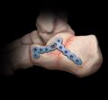
What Is Mortons Neuroma

Overview

intermetatarsal neuromaMorton?s Neuroma is a common foot condition characterized by pain and swelling in the ball of the foot, between the third and fourth toes. It?s caused by bones in your feet squeezing a nerve. Symptoms include a sharp, burning pain and possibly separation between the affected toes.

Causes

It’s not always clear what causes Morton’s neuroma, but several things seem to aggravate it. These include other foot-related problems and wearing restrictive footwear. It’s thought that Morton’s neuroma may be caused by the toe bones (metatarsal bones) pressing against the nerve when the gap between the bones is narrow. This causes the nerve and surrounding tissue to thicken.

Symptoms

While the condition may at first only appear during heavy repetitive stress or when wearing particular shoes which aggravate the foot, the Neuroma can become increasingly inflamed and produce more constant discomfort, lasting days or weeks. Runners may experience pain pushing off from starting blocks. Tight or narrow shoes as well as high heels likewise aggravate the Neuroma. A checklist of symptoms includes burning pain, occasionally numbness in the ball of the foot. Radiating pain from the ball of the foot to the toes. Intensifying pain during activity and when wearing shoes. Occasional numbness, discomfort, tingling or ?electrical shock sensation? in the toes. Pain between the third and fourth toes, often occurring from the outer side of one toe to the inner side of the adjoining toe. Pain upon leaving the starting blocks in running sports.

Diagnosis

Patients with classic Morton?s neuroma symptoms will have pain with pressure at the base of the involved toes (either between the 2nd and 3rd toes, or between the 3rd and 4th toes). In addition, squeezing the front of the foot together can exacerbate symptoms. As well, they may have numbness on the sides of one toe and the adjacent toe as this corresponds with the distribution of the involved nerve.

Non Surgical Treatment

Treatments may include rehabilitation measures to reduce nerve Irritation. Switching to low-heeled, wide-toed shoes with good arch support. Wearing padding in the shoes and/or between the toes. Wearing shoe inserts to correct a mechanical abnormality of the foot. Having ultrasound, electrical stimulation, whirlpool, and massage done on the foot. The foot may be injected with corticosteroids mixed with a local anesthetic in order to reduce pain. Relief may be only temporary, however, if the mechanical irritation is not also corrected. Injections with other types of medications such as alcohol, phenol, or vitamin B12 are sometimes used.

Surgical Treatment

Surgery for Morton’s neuroma is usually a treatment of last resort. It may be recommended if you have severe pain in your foot or if non-surgical treatments haven’t worked. Surgery is usually carried out under local anaesthetic, on an outpatient basis, which means you won’t need to stay in hospital overnight. The operation can take up to 30 minutes. The surgeon will make a small incision, either on the top of your foot or on the sole. They may try to increase the space around the nerve (nerve decompression) by removing some of the surrounding tissue, or they may remove the nerve completely (nerve resection). If the nerve is removed, the area between your toes may be permanently numb. After the procedure you’ll need to wear a special protective shoe until the affected area has healed sufficiently to wear normal footwear. It can take up to four weeks to make a full recovery. Most people (about 75%) who have surgery to treat Morton’s neuroma have positive results and their painful symptoms are relieved.

Prevention

To help reduce your chance of developing Morton’s neuroma avoid wearing tight and/or high-heeled shoes. Maintain or achieve ideal body weight. If you play sports, wear roomy, properly fitting athletic footwear.

The Way To Identify Inferior Calcaneal Spur

Inferior Calcaneal Spur

Overview

In the setting of plantar fasciitis, heel spurs are most often seen in middle-aged men and women, but can be found in all age groups. The heel spur itself is not thought to be the primary cause of pain, rather inflammation and irritation of the plantar fascia is thought to be the primary problem. A heel spur diagnosis is made when an x-ray shows a hook of bone protruding from the bottom of the foot at the point where the plantar fascia is attached to the heel bone.

Causes

Heel spurs develop as an abnormal growth in the heel bone due to calcium deposits that form when the plantar fascia pulls away from the heel. This stretching of the plantar fascia is usually the result of over-pronation (flat feet), but people with unusually high arches (pes cavus) can also develop heel spurs. Women have a significantly higher incidence of heel spurs due to the types of footwear often worn on a regular basis.

Inferior Calcaneal Spur

Symptoms

You may or may not experience any symptoms with your heel spurs. It is normally the irritation and inflammation felt in the tissues around your heel spur that cause discomfort. Heel pain is one of the first things you may notice, especially when pushing off the ball of your foot (stretches the plantar fascia). The pain can get worse over time and tends to be stronger in the morning, subsiding throughout the day; although it does return with increased activity. A sharp, poking pain in your heel that feels like you’re stepping on a stone can often be felt while standing or walking. You will sometimes be able to feel a bump on the bottom of your heel, and occasionally bruising may appear.

Diagnosis

Your doctor will discuss your medical history and will examine your foot and heel for any deformities and inflammation (swelling, redness, heat, pain). He/she will analyze your flexibility, stability, and gait (the way you walk). Occasionally an x-ray or blood tests (to rule out diseases or infections) may be requested.

Non Surgical Treatment

Ice and use arch support . If you can localize the spur, cut a hole in a pad of felt and lay the hole over the spur. This supports the area around the spur and reduces pressure on it. Massage the spur. Start gently with your thumb and gradually increase the pressure until you?re pushing hard directly on the spur with your knuckle or another firm object. Even it if hurts, it should help. Arch support. Build up an arch support system in your shoes. Try to equalize the pressure of your body weight throughout your arch and away from the plantar area. Use a ?cobra pad? or other device that supports the arch but releases pressure on the painful area. If homemade supports do not work, see a podiatrist about custom orthotics.

Surgical Treatment

In some cases, heel spurs are removed by surgery after an X-ray. While the surgery is typically effective, it?s a timely and expensive procedure. Even after surgery, heel spurs can re-form if the patient continues the lifestyle that led to the problem. These reasons are why most people who develop painful heel spurs begin looking for natural remedies for joint and bone pain. Surgery isn?t required to cure a heel spur. In fact, more than 90 percent of people get better with nonsurgical treatments. If nonsurgical methods fail to treat symptoms of heel spurs after 12 months, surgery may be necessary to alleviate pain and restore mobility.

Prevention

Walk around before you buy shoes. Before you purchase your shoes, do the following. Re-lace the shoes if you’re trying on athletic shoes. Start at the farthest eyelets and apply even pressure to the laces as you come closer to the tongue of the shoe. Make sure that you can wiggle your toes freely inside of the shoe. Also, make sure that you have at enough space between your tallest toe and the end of the shoe. You should have room equal to about the width of your thumb in the tip of your shoe. Walk around to make sure that the shoe has a firm grip on your heel without sliding up and down. Walk or run a few steps to make sure your shoes are comfortable. Shoes that fit properly require no break-in period.

How To Identify Calcaneal Spur

Heel Spur

Overview

A heel spur also known as a calcaneal spur, is a pointed bony outgrowth of the heel bone (calcaneus). Heel spurs do not always cause pain and often are discovered incidentally on X-rays taken for other problems. Heel spurs can occur at the back of the heel and also under the heel bone on the sole of the foot, where they may be associated with the painful foot condition plantar fasciitis.

Causes

When a patient has plantar fasciitis, the plantar fascia becomes inflamed and degenerative (worn out)–these abnormalities can make normal activities quite painful. Symptoms typically worsen early in the morning after sleep. At that time, the plantar fascia is tight so even simple movements stretch the contracted plantar fascia. As you begin to loosen the plantar fascia, the pain usually subsides, but often returns with prolonged standing or walking.

Inferior Calcaneal Spur

Symptoms

An individual with the lower legs turning inward, a condition called genu valgus or “knock knees,” can have a tendency toward excessive pronation. This can lead to a fallen arch and problems with the plantar fascia and heel spurs. Women tend to suffer from this condition more than men. Heel spurs can also result from an abnormally high arch. Other factors leading to heel spurs include a sudden increase in daily activities, an increase in weight, or a thinner cushion on the bottom of the heel due to old age. A significant increase in training intensity or duration may cause inflammation of the plantar fascia. High-heeled shoes, improperly fitted shoes, and shoes that are too flexible in the middle of the arch or bend before the toe joints will cause problems with the plantar fascia and possibly lead to heel spurs.

Diagnosis

The proper diagnosis of a heel spur often requires an X-ray. To make this process as convenient for his patients as possible, most clinics have an on-site digital X-ray and diagnostic ultrasound machines. This can make it unnecessary for patients to visit diagnostic imaging centers, allowing patients to receive more expedient treatment.

Non Surgical Treatment

There are many ways to treat heel spurs. Some remedies you can even do at home once a podiatrist shows you how. Heel spur treatment is very similar to treatment of plantar fasciitis. Here are a few of the most common treatments. First, your doctor will assess which activities are causing your symptoms and suggest rest and time off from these activities. Ice packs are used to control pain and reduce symptoms. Certain exercises and stretches help you to feel relief quickly. Medications that reduce inflammation and decrease pain are also used. Sometimes cortisone injections are given. Often special shoe orthotics can help to take the pressure off of the plantar fascia and reduce symptoms. Night splints that keep your heel stretched are sometimes recommended. Rarely, surgery is an option. A new treatment called extracorporeal shock wave therapy is being studied.

Surgical Treatment

Surgery involves releasing a part of the plantar fascia from its insertion in the heel bone, as well as removing the spur. Many times during the procedure, pinched nerves (neuromas), adding to the pain, are found and removed. Often, an inflamed sac of fluid call an accessory or adventitious bursa is found under the heel spur, and it is removed as well. Postoperative recovery is usually a slipper cast and minimal weight bearing for a period of 3-4 weeks. On some occasions, a removable short-leg walking boot is used or a below knee cast applied.

Bursitis Foot Pain Symptoms

Overview

Bursitis commonly affects joints used in repeated motions such as throwing a ball, or joints that bear pressure from being in the same position for a while, such as leaning on your elbows. The most common spots for bursitis are the shoulders, elbows or hips. Bursitis can also affect the knees (sometimes called ?housemaid?s knee? or ?vicar?s or preacher?s knee?), the heel of the foot or the base of the big toe. The good news is bursitis usually goes away with simple self-care treatments. However, not all cases of bursitis are from overuse, it can also be caused by an infection (called septic bursitis) or another condition such as arthritis. Therefore, it?s important to talk to your doctor if you think you have bursitis.

Causes

If the posterior-superior portion of the heel has an abnormally large bony prominence protruding from it (called a Haglund’s Deformity), in some instances it may rub against the Achilles Tendon. When this occurs, the bursa between the bone and the tendon will become inflamed, swollen, and painful. This condition is called Retrocalcaneal Bursitis. The presence of a Haglund’s Deformity does not insure that these problems will occur. In order for these problems to occur, the heel and foot must be tilted in such a way as to actually force this bony prominence into the bursa and tendon.

Symptoms

Unlike Achilles tendinitis, which tends to manifest itself slightly higher on the lower leg, Achilles tendon bursitis usually creates pain and irritation at the back of the heel. Possible signs of bursitis of the Achilles tendon include difficulty to rise on toes. Standing on your toes or wearing high heels may increase the heel pain. Inflammation and tenderness. The skin around your heel can become swollen and warm to the touch. Redness may be visible. Pain in the heel. Pain tends to become more prominent when walking, running, or touching the inflamed area. Stiffness. The back of your ankle may feel a little stiff due to the swelling of the bursa.

Diagnosis

A physical examination will be performed to determine if you have any signs of Achilles Bursitis or other ankle injury. He/she will look and feel the soft tissue and bones in your ankles to note any differences between the two of them. This will identify any abnormalities, such as swelling, bone deformities, atrophied muscles, redness and/or warmth on the skin. In many cases, the first sign that you have Achilles bursitis is swelling in the back of the foot and ankle pain.

Non Surgical Treatment

It is important to treat bursitis in the early stages to reduce the symptoms, minimize damage and maintain motion and strength in your foot. Resting your ankle, using proper cushioning, wearing comfortable footwear and reducing any activities that add pressure on your bursa will help to reduce your pain and bursitis inflammation.

Surgical Treatment

Bursectomy is a surgical procedure used to remove an inflamed or infected bursa, which is a fluid-filled sac that reduces friction between tissues of the body. Because retrocalcaneal bursitis can cause chronic inflammation, pain and discomfort, bursectomy may be used as a treatment for the condition when it is persistent and cannot be relived with other treatments. During this procedure, a surgeon makes small incisions so that a camera may be inserted into the joint. This camera is called an arthroscope. Another small incision is made so that surgical instruments can be inserted to remove the inflamed bursa.

Prevention

Continue to wear your orthotics for work and exercise to provide stability and restore foot function. Select suitable shoes for work and physical activity that provide stability for the heel. Regular stretching of the calf muscle can prevent heel bursitis. Do not suddenly increase activity amount without appropriate conditioning.

Nonsurgical Hammer Toe Repair

HammertoeOverview

A hammertoe is a toe that is contracted at the PIP joint (middle joint in the toe), potentially leading to severe pressure and pain. Ligaments and tendons that have tightened cause the toe’s joints to curl downwards. Hammer toes may occur in any toe except the big toe. There is often discomfort at the top part of the toe due to rubbing against the shoe.

Causes

Hammer toe results from shoes that don’t fit properly or a muscle imbalance, usually in combination with one or more other factors. Muscles work in pairs to straighten and bend the toes. If the toe is bent and held in one position long enough, the muscles tighten and cannot stretch out. Shoes that narrow toward the toe may make your forefoot look smaller. But they also push the smaller toes into a flexed (bent) position. The toes rub against the shoe, leading to the formation of corns and calluses, which further aggravate the condition. A higher heel forces the foot down and squishes the toes against the shoe, increasing the pressure and the bend in the toe. Eventually, the toe muscles become unable to straighten the toe, even when there is no confining shoe.

HammertoeSymptoms

The symptoms of a hammer toe are usually first noticed when a corn develops on the top of the toe and becomes painful, usually when wearing tight shoes. There may be a bursa under the corn or instead of a corn, depending on the pressure. Most of the symptoms are due to pressure from footwear on the toe. There may be a callus under the metatarsal head at the base of the toe. Initially a hammer toe is usually flexible, but when longstanding it becomes more rigid.

Diagnosis

Hammer toes Hammer toes may be easily detected through observation. The malformation of the person’s toes begin as mild distortions, yet may worsen over time – especially if the factors causing the hammer toes are not eased or removed. If the condition is paid attention to early enough, the person’s toes may not be permanently damaged and may be treated without having to receive surgical intervention. If the person’s toes remain untreated for too long, however the muscles within the toes might stiffen even more and will require invasive procedures to correct the deformity.

Non Surgical Treatment

Prescription strength medicines to decrease pain and inflammation. Physical Therapy. To strengthen poorly functioning muscles and stretch tight muscles that may be exacerbating the toes. Special ultrasound techniques may reduce inflammation. Custom Foot Orthotics. An orthotic with an exact mold of your foot to better align and support the foot to ease current discomfort and prevent future progression. Toe Splints or Pads. Specific pads may prevent pressure and physical irritation in shoes. Toe splints and toe spacers physically realign the toes and can lessen pain and halt or stall hammer toe progression. Cortisone injections are strong anti-inflammatory agents to decrease pain, and swelling directly at the toe region. Injections only treat the symptoms, and in some cases used in caution (and sparingly) they can weaken supporting ligaments of the toe(s).

Surgical Treatment

Sometimes when the joints are removed the two bones become one as they are fused in a straightened position. Many times one toe will be longer than another and a piece of bone is removed to bring the toes in a more normal length in relation to each other. Sometimes tendons will be lengthened, or soft tissue around the joints will be cut or rebalanced to fix the deformity. Angular corrections may also be needed. The surgeon may place fixation in your foot as it heals which may include a pin, or wires.

Hammer ToePrevention

Some tips on how to avoid getting hammertoes when wearing high heels has to do with the shoes that you choose and what you do to your feet after you wear the high heels. A closed toe shoe like a stiletto pump is going to put more pressure on the front of the foot, forcing the toes to contract in and start forming the hammertoes. Women who start having hammertoes usually complain about pain in their feet when they wear the closed shoe, usually in the winter season, and when in the warmer weather and they wear the open-toed shoes, there?s much less pressure on the front of the foot and they experience much less pain.

Hammer Toe Disorder

Hammer ToeOverview

Hammertoes are quite common and may range from mild to severe. A hammertoe is a contracture, or bending, of one or more toes, usually due to an imbalance between the muscles or tendons on the top and bottom of the toes. Over time, the affected toes lose flexibility, becoming rigid and fixed in a contracted position. The abnormal bend positions the knuckle of the toe upward, causing it to push against the top of the shoe leading to additional problems. The condition usually becomes progressively worse if not treated.

Causes

Hammertoes are usually structural in nature. Many times this is the foot structure you were born with and other factors have now made it so that symptoms appear. The muscles in your foot may become unbalanced over time, allowing for a deformity of the small bones in each toe. With longstanding deformity the toe may become rigid. Sometimes one toe is longer than another and this causes a buckling of the digit. A hammertoe may also be caused by other foot deformities such as a bunion. Trauma or other surgery of your foot may predispose you to having the condition if your foot structure is altered.

HammertoeSymptoms

Here is a look at some of the symptoms hammertoe can cause. They include hammer-like or claw-like appearance of the toe. Pain when walking or moving the foot. Difficulty moving the toe. Corns may form on top of the toe. Callus may form on the sole of the foot. During the initial stages, you may be able to manually straighten your toe. This is called a flexible hammertoe. But as time passes, the toe will not move as easily and will continue to look like a hammer. Pressure and irritation over the joint can cause a blister to develop and become a corn over time. These corns have the potential to become infected and cause additional symptoms such as redness, bleeding, and difficulty wearing shoes and socks. Corns are the main cause of pain when hammertoes are developing.

Diagnosis

Most health care professionals can diagnose hammertoe simply by examining your toes and feet. X-rays of the feet are not needed to diagnose hammertoe, but they may be useful to look for signs of some types of arthritis (such as rheumatoid arthritis) or other disorders that can cause hammertoe. If the deformed toe is very painful, your doctor may recommend that you have a fluid sample withdrawn from the joint with a needle so the fluid can be checked for signs of infection or gout (arthritis from crystal deposits).

Non Surgical Treatment

The most common treatment is to wear more comfortable shoes. When choosing a shoe, make sure the toe area is high and broad and has enough room for hammer toes. If there is chronic pain, surgery may be needed to correct a malalignment. Surgical treatments are Hammer toe aimed at loosening up the contracted toe joints to allow them to align properly. Other types of treatment are products designed to relieve hammer toes, such as hammer toe crests and hammer toe splints. These devices will help hold down the hammer toe and provide relief to the forefoot. Gel toe shields and gel toe caps can also be used. Gel toe shields and toe caps will help eliminate friction between the shoe and the toe, while providing comfort and lubrication.

Surgical Treatment

If this fails or if treatment is not sought until the toes are permanently misaligned, then surgery may be required. Surgery may involve either cutting the tendon or fusing the joint. Congenital conditions should be treated in early childhood with manipulations and splinting.

Hammer ToePrevention

Preventing foot problems, including hammertoes, is often a matter of wearing the right shoes and taking care of your feet. Check your feet regularly for problems. This is especially true if you have diabetes or any other medical condition that causes poor circulation or numbness in your toes. If you do, check feet daily so that problems can be caught early on.

Bunions Causes And Treatments

Overview
Bunions
A bunion is an enlargement of bone at the great toe joint. Tight shoes don’t cause bunions, but they can aggravate them. Bunions are often inherited and become worse over time if left untreated they can cause pain, swelling, skin irritation and other foot problems. Bunion deformities are often part of a more generalized problem related to improper foot motion. There are presently over 25 types of bunion procedures performed today. The choice of procedure is based on many factors.

Causes
Despite the popular belief, wearing high heels and too-narrow shoes does not cause bunions. Wearing them can irritate, aggravate, or accelerate the formation of the bunion, but are not the root cause. Bunions are more commonly inherited, if your parents or grandparents had bunions, you may also get one. Bunions can also be caused by trauma or injury to the joints, ligaments, or bones of the foot.
SymptomsNo matter what stage your bunion is in, you can be in pain. Though bunions take years to develop, you can experience pain at any stage. Some people don?t have bunion pain at all. Pain from a bunion can be severe enough to keep you from walking comfortably in normal shoes. The skin and deeper tissue around the bunion also may become swollen or inflamed.

Diagnosis
Your doctor is very likely to be able to diagnose your bunion simply by examining your foot. Even before that, he or she will probably ask about your family and personal medical history and evaluate the types of shoes you wear. You’ll be asked about your symptoms, when they started and when they occur. You may also be asked to flex your toe so that your doctor can get an idea of your range of motion. He or she may order x-rays in order to determine the extent of your deformity.

Non Surgical Treatment
Fortunately, many bunions never go on to cause problems other than the cosmetic appearance. The easiest option is to try different shoes or padding, however this is not the answer for everyone. The various straps and braces that are commercially available are not proven to be particularly effective.
Bunion Pain

Surgical Treatment
Some sufferers choose to have the bunion surgically removed. This should always be a last resort as all surgeries carry risks. There are several types of surgical procedures to remove bunions and before deciding, you should speak to your surgeon at length about the facts and risks associated with surgery, including the recovery time and success rate of the operation to be done. Please note that if you have a surgical procedure and then return to your high heels and narrow-toed shoes, the bunion is likely to reoccur.

What Causes Feet To Over Pronate

Overview

When sitting, an over-pronating foot appears quite normal, i.e. showing a normal arch with room under the underside of the foot. The moment you get up and put weight on your feet the situation changes: the arches lower and the ankle slightly turns inwards. When you walk or run more weight is placed on the feet compared to standing and over-pronation will become more evident. When walking barefoot on tiles or timber floors over-pronation is more visible, compared to walking on carpet or grass.Foot Pronation

Causes

Congenital “Flat Feet” – an individual may be born with feet that lack an appropriately supportive arch thereby predisposing the individual to this foot condition. Excessive Weight (Obesity) Too much weight on the foot from either obesity or pregnancy may be a factor. Repetitive Impact walking on flat, hard surfaces continuously places unnatural stress on the foot arch.

Symptoms

Common conditions that develop with prolonged overpronation typically include plantar fasciitis, achilles tendonitis, shin splints, posterior tibial stress syndrome and even IT band syndrome. With long term neglect you may see the development of bunyons, foot deformities and early onset of hip and knee arthritis.

Diagnosis

The best way to discover whether you have a normal gait, or if you overpronate, is to visit a specialty run shop, an exercise physiologist, a podiatrist or a physical therapist who specializes in working with athletes. A professional can analyze your gait, by watching you either walk or run, preferably on a treadmill. Some facilities can videotape your gait, then analyze the movement of your feet in slow-motion. Another (and less costly) way is to look at the bottom of an older pair of run shoes. Check the wear pattern. A person with a normal gait will generally see wear evenly across the heel and front of the shoe. A person who overpronates will likely see more wear on the OUTside of the heel and more wear on the INside of the forefoot (at the ball). A person who supinates will see wear all along the outer edges of the shoe. You can also learn about your gait by looking at your arches. Look at the shape your wet feet leave on a piece of paper or a flat walking surface.Foot Pronation

Non Surgical Treatment

Orthotics are medical devices used to provide support to correct a physical abnormality. They can provide arch support when needed to remedy over-pronation, and in this particular cases the orthoses used are usually convenient shoe inserts. These can be taken in and out of shoes, and will be carefully tailored by your podiatrist to the specifics of your foot. It can take some weeks before the effects of the inserts can become truly noticeable, and in many cases your podiatrist will want to review your orthotics within a few weeks to make fine adjustments based on how well they have worked to reduce your pain.

Surgical Treatment

The MBA implant is small titanium device that is inserted surgically into a small opening between the bones in the hind-mid foot: the talus (ankle bone) and the calcaneus (heel bone). The implant was developed to help restore the arch by acting as a mechanical block that prevents the foot from rolling-in (pronation). In the medical literature, the success rate for relief of pain is about 65-70%. Unfortunately, about 40% of people require surgical removal of the implant due to pain.

Will Severs Disease Require Surgical Treatments?

Overview

A common cause of heel pain in growing adolescents, particularly those that are actively participating in sport is a condition known as Severs disease. While the suggestion of a ?disease? to your child may conjure up images of a life threatening disorder crippling your child, Severs disease is not nearly so sinister and can be easily fixed.

Causes

Inflammation occurs at the insertion of the achilles tendon into the back of the heel due to a number of reasons. One or several of the following may cause the initiation of Sever?s disease. Rapid growth spurt. Tight calf muscles. Change in footwear (soccer boots / athletic shoes no heel). Excessive rolling in of feet. Poor warm up routine. Remember this condition usually settles as the growth plate fuses within 6-12 months.

Symptoms

The main symptom of sever’s disease is pain and tenderness at the back of the heel which is made worse with physical activity. Tenderness will be felt especially if you press in or give the back of the heel a squeeze from the sides. There may be a lump over the painful area. Another sign is tight calf muscles resulting with reduced range of motion at the ankle. Pain may go away after a period of rest from sporting activities only to return when the young person goes back to training.

Diagnosis

Sever?s disease can be diagnosed based on your history and symptoms. Clinically, your physiotherapist will perform a “squeeze test” and some other tests to confirm the diagnosis. Some children suffer Sever?s disease even though they do less exercise than other. This indicates that it is not just training volume that is at play. Foot and leg biomechanics are a predisposing factor. The main factors thought to predispose a child to Sever?s disease include decrease ankle dorsiflexion, abnormal hind foot motion eg overpronation or supination, tight calf muscles, excessive weight-bearing activities eg running.

Non Surgical Treatment

The aims are to reduce trauma to the heel, allow rest/recovery and prevent recurrence. Most cases are successfully treated using physiotherapy and exercises, eg to stretch the gastrocnemius-soleus complex, to mobilise the ankle mortise, subtalar joint and medial forefoot. Soft orthotics or heel cups. Advice on suitable athletic footwear. Other modes of treatment are in severe cases, temporarily limiting activity such as running and jumping. Ice and non-steroidal anti-inflammatory drugs (NSAIDs), which can reduce pain. In very severe cases, a short period of immobilisation (eg 2-3 weeks in a case in mild equinus position) has been suggested.

Surgical Treatment

The surgeon may select one or more of the following options to treat calcaneal apophysitis. Reduce activity. The child needs to reduce or stop any activity that causes pain. Support the heel. Temporary shoe inserts or custom orthotic devices may provide support for the heel. Medications. Nonsteroidal anti-inflammatory drugs (NSAIDs), such as ibuprofen, help reduce the pain and inflammation. Physical therapy. Stretching or physical therapy modalities are sometimes used to promote healing of the inflamed issue. Immobilization. In some severe cases of pediatric heel pain, a cast may be used to promote healing while keeping the foot and ankle totally immobile. Often heel pain in children returns after it has been treated because the heel bone is still growing. Recurrence of heel pain may be a sign of calcaneal apophysitis, or it may indicate a different problem. If your child has a repeat bout of heel pain, be sure to make an appointment with your foot and ankle surgeon.

Posterior Tibial Tendon Dysfunction

Overview
There are four stages of posterior tibial tendon dysfunction. In the first stage the posterior tibial tendon is inflamed but has normal strength. There is little to no change in the arch of the foot. In stage two the tendon is partially torn or shows degenerative changes and as a result loses strength. There is considerable flattening of the arch without arthritic changes in the foot. Stage three results when the posterior tibial tendon is torn and not functioning. As a result the arch is completely collapsed with arthritic changes in the foot. Stage four is identical to stage three except that the ankle joint also becomes arthritic.
Flat Foot

Causes
The posterior tibial tendon, which connects the bones inside the foot to the calf, is responsible for supporting the foot during movement and holding up the arch. Gradual stretching and tearing of the posterior tibial tendon can cause failure of the ligaments in the arch. Without support, the bones in the feet fall out of normal position, rolling the foot inward. The foot’s arch will collapse completely over time, resulting in adult acquired flatfoot. The ligaments and tendons holding up the arch can lose elasticity and strength as a result of aging. Obesity, diabetes, and hypertension can increase the risk of developing this condition. Adult acquired flatfoot is seen more often in women than in men and in those 40 or older.

Symptoms
Patients often experience pain and/or deformity at the ankle or hindfoot. When the posterior tibial tendon does not work properly, a number of changes can occur to the foot and ankle. In the earlier stages, symptoms often include pain and tenderness along the posterior tibial tendon behind the inside of the ankle. As the tendon progressively fails, deformity of the foot and ankle may occur. This deformity can include progressive flattening of the arch, shifting of the heel so that it no longer is aligned underneath the rest of the leg, rotation and deformity of the forefoot, tightening of the heel cord, development of arthritis, and deformity of the ankle joint. At certain stages of this disorder, pain may shift from the inside to the outside aspect of the ankle as the heel shifts outward and structures are pinched laterally.

Diagnosis
Diagnostic testing is often used to diagnose the condition and help determine the stage of the disease. The most common test done in the office setting are weightbearing X-rays of the foot and ankle. These assess joint alignment and osteoarthritis. If tendon tearing or rupture is suspected, the gold standard test would be MRI. The MRI is used to check the tendon, surrounding ligament structures and the midfoot and hindfoot joints. An MRI is essential if surgery is being considered.

Non surgical Treatment
Nonoperative treatment of posterior tibial tendon dysfunction can be successful with the Arizona AFO brace, particularly when treatment is initiated in the early stages of the disease. This mandates that the orthopedist has a high index of suspicion when evaluating patients to make an accurate diagnosis. Although there is a role for surgical management of acquired flat feet, a well-fitted, custom-molded leather and polypropylene orthosis can be effective at relieving symptoms and either obviating or delaying any surgical intervention. In today’s climate of patient satisfaction directed health care, a less invasive treatment modality that relieves pain may prove to be more valuable than similar pain relief that is obtained after surgery. Questions regarding the long-term results of bracing remain unanswered. Future studies are needed to determine if disease progression and arthrosis occur despite symptomatic relief with a brace. Furthermore, age- and disease stage-matched control groups who are randomized to undergo surgery or bracing are necessary to compare these different treatment modalities.
Acquired Flat Feet

Surgical Treatment
Good to excellent results for more than 80% of patients have been reported at five years’ follow up for the surgical interventions recommended below. However, the postoperative recovery is a lengthy process, and most surgical procedures require patients to wear a plaster cast for two to three months. Although many patients report that their function is well improved by six months, in our experience a year is required to recover truly and gain full functional improvement after the surgery. Clearly, some patients are not candidates for such major reconstructive surgery.